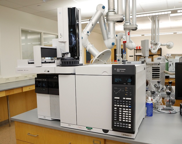
GC/MS is considered "state of the art" technology for drug identification. In basic terms, the instrument consists of a precisely regulated oven housing a long hollow coil or "column" (the GC) attached to a molecular weight detector (the MS). The hollow column used in our work is typically 20 meters long (approximately 60 feet) and has an inside diameter of 0.18 millimeters. For reference, 0.18 mm is approximately 1/14,000 of an inch!
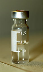
For GC/MS, a sample is dissolved in an appropriate solvent and injected into the end of the column called the "injection port" of the GC. The injection port is typically heated to over 200 degrees centigrade. This heat vaporizes the sample (changes it from liquid to gas) and allows the molecules to migrate through the column under controlled conditions.
The most important factor with GC is that compounds will migrate through the column at different rates. This principle allows a complex mixture of compounds to be separated into individual components or "peaks". A simple analogy to this characteristic is a horse track. All horses in a typical marathon start the race at the same time, but based on the attributes of each horse, the participants will cross the finish line at different times!
The length of time required for a molecule to pass through the entire column is based on its physical and chemical characteristics (which are unique to that molecule) and is referred to the retention time.
The far end of the column is attached to the Mass Spectrometer. After the molecules are sorted by retention time, they exit the column and enter the MS where the separated compound is bombarded by a narrow beam of electrons. This beam shatters the molecules into characteristic fragments. The instrument is always "tuned" to highly specific settings, therefore these molecules always break apart into the same pieces.
Within the mass spectrometer is a "mass filter" which uses electromagnetic fields to focus and sort the fragments by molecular weight. The end result is a mass spectrum (a type of bar chart) that displays the amount of each fragment and its corresponding molecular weight. This bar chart forms a specific "molecular fingerprint" for the compound in question. By comparing this mass spectrum (and the retention time) to certified standards and/or a vast drug reference library, the Forensic Chemist can make a positive identification of the drug.